Chapter 1 of 5 - Histology Course
Introduction to Histology & Tissue Preparation
Histology is how we see structure at the tissue scale. This chapter covers fixation, the paraffin workflow, routine H&E staining, how to choose magnification, and how to interpret basophilic versus eosinophilic patterns on a slide.
What Is Histology?
Histology (microscopic anatomy) examines how cells assemble into tissues and how tissues form organs. Pathologists and researchers rely on thin sections and stains because many diseases first alter tissue architecture long before gross anatomy changes. Students use the same methods to connect lecture concepts to real morphology.
Standard light microscopy uses sections typically 4-7 micrometers thick in paraffin, stained so nuclei, cytoplasm, and matrix separate visually. The most common routine stain worldwide is hematoxylin and eosin (H&E). Special stains and immunohistochemistry add specificity when H&E alone is not enough.
"Histology is the bridge between the cell and the organ."
- common teaching line in microscopic anatomy
The Four Primary Tissue Types
Almost every organ maps back to combinations of these four categories. On exams, you are often asked to identify the dominant pattern in a field of view and justify it using nucleus shape, matrix abundance, striations, or layering.
Epithelial
Sheets, glands, polarity
Connective
Matrix, fibers, cells
Muscle
Skeletal, cardiac, smooth
Nervous
Neurons, glia, layers
Paraffin Section Workflow (Overview)
Teaching labs and clinical labs both follow the same logical sequence. Each step exists to preserve chemistry, harden tissue for thin cutting, or restore water compatibility before dyes bind predictably.
Fixation
Cross-link proteins, stop autolysis
Dehydration - Clearing - Infiltration
Alcohol series, xylene or substitute, molten paraffin
Embedding & sectioning
Paraffin block, microtome ribbons, float on water bath
Mount, dewax, rehydrate, stain
H&E or protocol-specific sequence
Frozen sections shorten the path for intraoperative consultation: tissue is frozen and cut without full paraffin embedding. Cell detail and architecture can differ from permanent sections, so pathologists interpret frozen results with that limitation in mind.
H&E: Basophilic vs Eosinophilic Patterns
Staining language shows up constantly in lab manuals and board-style questions. Think "acid-base" chemistry: hematoxylin is basic and binds acidic structures (hence nuclei rich in nucleic acids stain basophilic). Eosin is acidic and binds basic proteins that predominate in much of the cytoplasm and in collagen.
| Appearance | Typical structures | Why (simplified) |
|---|---|---|
| Basophilic (blue-purple) | Nuclei, rough ER-rich cytoplasm, some matrix | Hematoxylin binds acidic phosphate and carboxyl groups |
| Eosinophilic (pink) | Most cytoplasm, collagen, RBCs | Eosin binds many basic protein side chains |
| Clear or washed out | Large lipid droplets, some mucins | Lipid dissolved in processing; mucin may need special stains |
Magnification Strategy on a Glass Slide
Start at low power to judge overall architecture: Is this epithelium versus connective tissue dominant? Is there a lumen or surface? Are layers organized normally? Then move to high power for nuclear detail, mitoses, inflammation, or organisms.
If you begin at maximum magnification, you may miss the forest for the trees. Exam questions often show a field that only makes sense once you infer organ context from low-power pattern recognition.
Formaldehyde (fixation)
formaldehyde
Formaldehyde (often from formalin solution) cross-links proteins in tissue, preserving structure for embedding and sectioning. It is a foundational step before most paraffin histology.
Formula
CH2O
Mol. Weight
30.03 g/mol
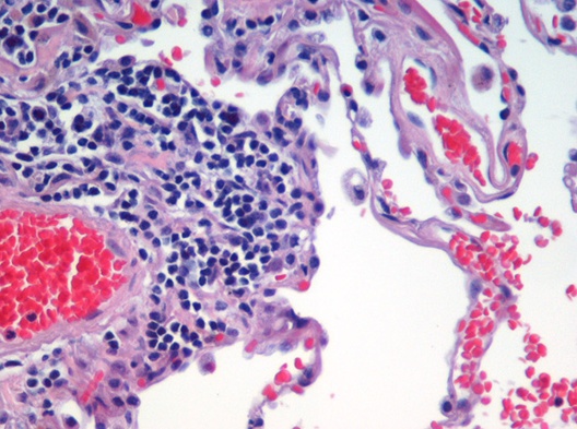
H&E-stained lung tissue: nuclei appear blue-purple, cytoplasm and matrix pink, and air spaces relatively clear. This illustrates the contrast H&E provides for teaching histology.
Wars, Wikimedia Commons, CC BY 2.0
Why Fixation Quality Matters
Under-fixed tissue autolyzes; over-fixation can mask antigens needed for immunostains. Clinicians and biobank protocols therefore standardize time in fixative and specimen handling. For course exams, know that fixation is the first domino: it influences shrinkage, antigenicity, and how crisply nuclei and membranes appear.
Quick Check
In routine H&E sections, which structure is expected to stain most basophilic (blue-purple) with hematoxylin?
Fill in the Blank
The standard pair of dyes used in most diagnostic histology labs is hematoxylin and________.
Quick Check
Which order best describes a rational light-microscopy approach to an H&E slide you have never seen?
What You Will Learn in This Course
This five-chapter histology course walks from preparation and staining through each major tissue class. Each page includes quick checks, fill-in prompts, and links to trusted figures. Finish with the histology game and the study guide for consolidation.
- Introduction (this page) - workflow, H&E logic, magnification
- Epithelial tissue - classification, glands, basement membrane cues
- Connective tissue - matrix, fiber patterns, adipose
- Muscle and nervous tissue - striations, intercalated discs, CNS layering
- Stains and laboratory methods - eosin chemistry, artifacts, special stains overview
Was this helpful? Rate it!
Create a free Lorea account
Turn your notes and PDFs into full AI-generated courses with quizzes, summaries, flashcards, and narrated videos — then publish, download, and share them however you like.