Chapter 5 of 5 - Anatomy Course
Head, Neck & Neuroanatomy
The skull protects the brain and houses special senses. Cranial nerves exit through named foramina; the neck carries major vessels and the airway from thorax to cranial base.
Integrated questions link pupil size, gaze palsies, and facial weakness to segmental anatomy - know the skull base map and brainstem crossings.
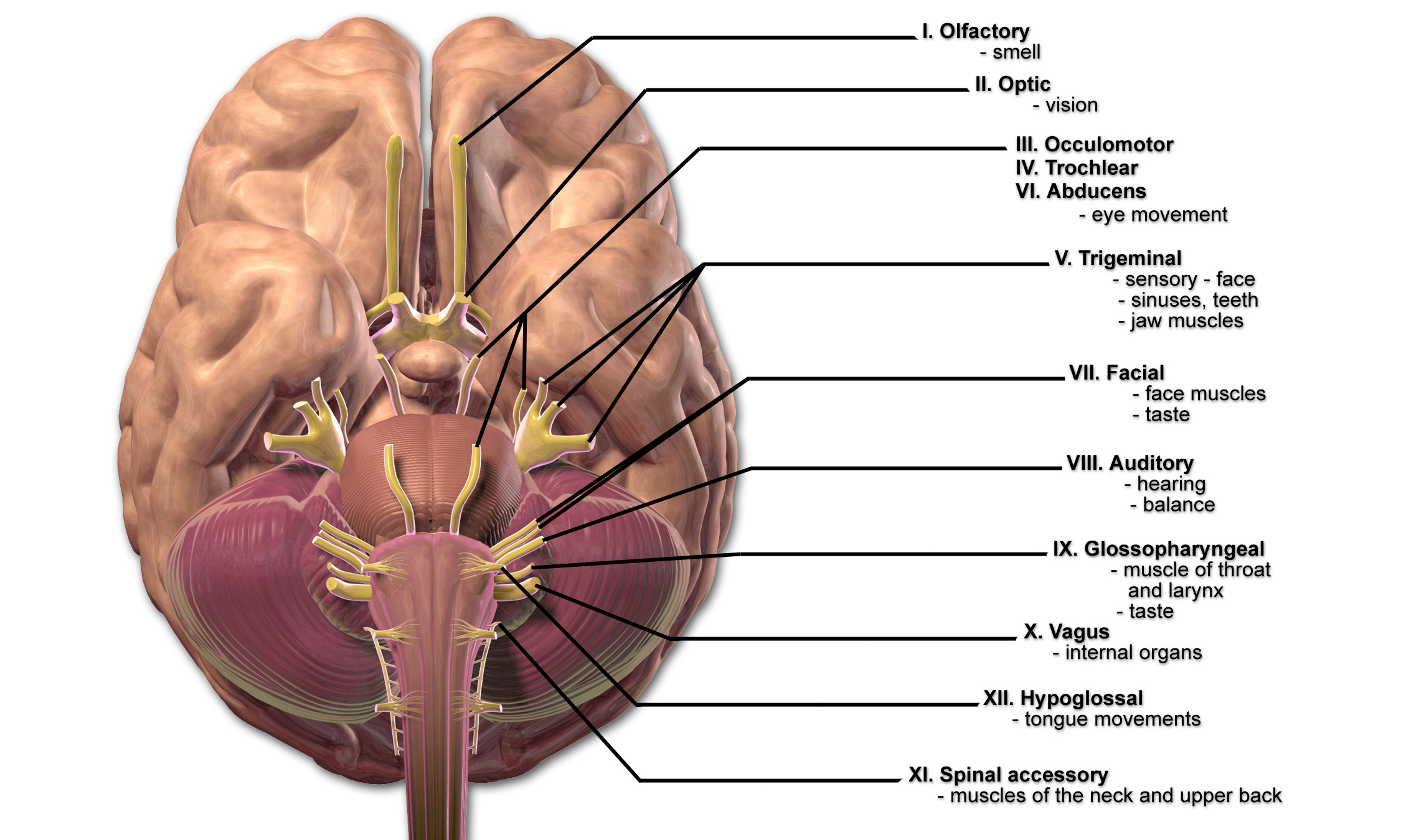
Skull Base and Foramina
Each cranial nerve has a predictable course: optic (II) through the optic canal, maxillary (V2) via foramen rotundum, mandibular (V3) via foramen ovale, and so on. Masses, fractures, and infiltrative processes produce syndromes by structure at each opening.
Smell (I)
Olfactory filaments - trauma, meningioma patterns.
Vision (II)
Optic nerve, chiasm, tract - field cuts.
Eye movements (III, IV, VI)
Ptosis, diplopia, strabismus localizations.
Face sensation & motor (V, VII)
Trigeminal divisions; facial nerve motor branches.
Memorize cranial nerve numbers and functions first; then attach each to skull base exit and clinical deficit patterns.
Acetylcholine
Cholinergic synapses at the neuromuscular junction and parasympathetic terminals tie molecular signaling to the motor and autonomic patterns you localize on exam.
Formula
C7H16NO2
Mol. Weight
145.21 g/mol
Meninges and Venous Sinuses
The dura mater has periosteal and meningeal layers; arachnoid bridges subarachnoid CSF spaces; pia adheres to brain. Epidural versus subdural bleeds differ by vessel source and bridging vein involvement - classic board contrast.
| Space | Key idea |
|---|---|
| Subarachnoid | CSF, circle of Willis, aneurysmal rupture patterns |
| Epidural | Arterial (middle meningeal) lens-shaped on imaging classically |
| Subdural | Bridging veins; crescentic collection; elderly, alcohol use |
Quick Check
Which cranial nerve carries parasympathetic fibers to the pupil sphincter and ciliary body?
Fill in the Blank
The meningeal layer directly adherent to the surface of the brain is the________ mater.
Upper versus lower motor neuron signs
Tone, reflexes, atrophy patterns.
Crossed versus uncrossed deficits
Brainstem lesions often show ipsilateral face, contralateral body.
Map to artery or nerve
MCA stroke versus peripheral VII palsy.
Was this helpful? Rate it!
Create a free Lorea account
Turn your notes and PDFs into full AI-generated courses with quizzes, summaries, flashcards, and narrated videos — then publish, download, and share them however you like.