Apical, basal and lateral: the anatomy of cell polarity
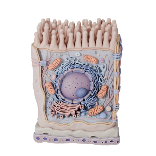
An epithelial cell has three distinct territories. The apical membrane faces the lumen or the outside world. The basal membrane sits on the basement membrane, anchored to the connective tissue underneath. The lateral membranes contact neighbouring cells. These are not just positional labels — each domain carries a different set of membrane proteins. Sodium-potassium ATPase pumps sit almost exclusively in the basolateral membrane, while specific nutrient transporters and channels sit apically. That separation is what makes transport directional: a glucose molecule can only travel lumen-to-blood, never the reverse, because the machinery for each step faces a different way. Lose the polarity and the tissue stops functioning as a barrier, which is one of the early hallmarks of carcinoma.
Microvilli and the brush border
The dense fringe on the apical surface of the model is microvilli — finger-like projections roughly one micrometre tall, each built around a core bundle of actin filaments cross-linked by villin and fimbrin and anchored into a mesh called the terminal web. Their only purpose is surface area. In the small intestine, microvilli multiply the absorptive surface by around twenty-fold on top of the amplification already provided by villi and folds. The membrane covering them is studded with brush border enzymes — lactase, sucrase-isomaltase, alkaline phosphatase — so final-stage digestion happens on the cell surface, millimetres from where absorption occurs. Microvilli are frequently confused with cilia, but they are structurally unrelated: microvilli are actin-based and immotile, cilia are microtubule-based with a 9+2 arrangement and beat actively.
Four junctions, four different jobs
Epithelial cells are stitched to each other by a stack of junctional complexes, and exam questions almost always turn on telling them apart. The tight junction (zonula occludens) sits most apically and is built from claudins and occludin; it seals the space between cells so material cannot leak around them, and it also acts as a fence that stops apical and basolateral membrane proteins from mixing — so the tight junction is what physically maintains polarity. Below it, the adherens junction uses E-cadherin linked to the actin cytoskeleton. Desmosomes use desmoglein and desmocollin tied to keratin intermediate filaments, and they exist purely for mechanical strength, which is why they are densest in tissue that gets stretched and abraded. Gap junctions are different in kind: connexon channels that directly couple the cytoplasm of adjacent cells, letting ions and molecules under about one kilodalton pass between them. Hemidesmosomes anchor the basal surface downward to the basement membrane using integrins rather than cadherins.
The basement membrane, and why epithelium has no blood supply
Underneath every epithelium is a thin sheet of extracellular matrix: the basal lamina, made of laminin, collagen IV, nidogen and perlecan, backed by a reticular lamina of collagen III. Together they form the basement membrane visible as the layered base of the 3D model. Critically, epithelium is avascular — no capillary ever enters it. Every nutrient and every oxygen molecule has to diffuse across the basement membrane from vessels in the connective tissue below. This one constraint explains a surprising amount of histology: it caps how thick an epithelium can get, it is why stratified squamous epithelium becomes progressively flatter and eventually dies as cells are pushed away from their supply, and it is why breaching the basement membrane is the definition of invasive rather than in-situ carcinoma.
Why these cells are so densely packed with mitochondria
Active transport is metabolically brutal. Sodium-potassium ATPase alone can consume a large fraction of a cell's total ATP budget, and epithelial cells specialised for transport run it continuously to maintain the gradients that every secondary transporter depends on. That is why the mitochondrial density in the model is high and concentrated toward the basal region, near the pumps that need the ATP. The same logic scales across the body: proximal tubule cells in the kidney and enterocytes in the small intestine are among the most mitochondria-rich cells you will find, purely because of the transport load they carry.
Classification: shape times layers
Epithelia are named on two axes simultaneously. Layers give simple (one cell thick), stratified (multiple layers), or pseudostratified (one layer that looks like several because nuclei sit at different heights). Shape gives squamous (flat), cuboidal (roughly square), or columnar (tall). Combine them and you get the standard set: simple squamous for gas exchange in alveoli and filtration in glomeruli, simple cuboidal in kidney tubules and glands, simple columnar with microvilli lining the gut, pseudostratified ciliated columnar in the airways, and stratified squamous where abrasion resistance matters — skin, oesophagus, vagina. Transitional epithelium (urothelium) is the outlier, changing shape as the bladder fills.
What to look for in the 3D model
Start by rotating the model so you are looking edge-on rather than from above; the polarity only becomes obvious in profile. Find the dense apical fringe of microvilli, then trace down the lateral surface to see where neighbouring cells would seal against it. Zoom into the base to see the layered cutaway of the basement membrane, and note how the nucleus sits basally rather than centrally — a consistent feature of tall absorptive epithelium that shows up constantly in histology slides.