The cell wall: rigidity without a skeleton
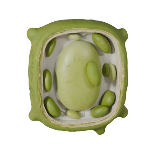
Outside the plasma membrane sits a wall built from cellulose microfibrils embedded in a matrix of hemicellulose and pectin. Adjacent cells are glued together by a shared pectin-rich layer called the middle lamella. Young cells have a flexible primary wall that can still expand; cells that have finished growing may add a rigid secondary wall inside it, often reinforced with lignin, which is essentially what wood is. The wall does two jobs at once. It provides mechanical support, letting a plant stand upright without bones. And it acts as a pressure vessel — because the wall resists expansion, a plant cell placed in pure water does not burst the way an animal cell would. It swells until wall pressure balances water entry and then stops.
The central vacuole and turgor pressure
The single largest feature in the model is the central vacuole, which in a mature plant cell can occupy eighty to ninety percent of the cell's volume, pressing the cytoplasm into a thin layer against the wall. It is bounded by a membrane called the tonoplast and filled with water, dissolved ions, sugars, pigments and metabolic waste. Its most important mechanical role is generating turgor pressure: water drawn in osmotically pushes outward against the wall, and that pressure is what keeps non-woody tissue firm. Put the cell in a hypertonic solution and water leaves, the vacuole shrinks, the membrane pulls away from the wall — plasmolysis — and at the whole-plant scale you see wilting. Watering a wilted plant restores turgor within hours, which is the same phenomenon running in reverse.
Chloroplasts: where photosynthesis actually happens
Chloroplasts are double-membraned organelles containing a third internal membrane system: flattened sacs called thylakoids, stacked into columns known as grana, suspended in a fluid stroma. The division of labour between these compartments is a standard exam point. The light-dependent reactions happen in the thylakoid membranes, where chlorophyll harvests photons, water is split, and a proton gradient across the thylakoid membrane drives ATP synthesis. The Calvin cycle — carbon fixation proper, via the enzyme RuBisCO — happens in the stroma outside the thylakoids. Chloroplasts also carry their own small circular genome and their own ribosomes, and they divide independently of the cell, all of which is evidence for the endosymbiotic theory that they descend from free-living photosynthetic bacteria.
Plasmodesmata and the symplast
Because plant cells are walled off from each other, they need dedicated channels to communicate. Plasmodesmata are narrow pores threading through the walls between neighbouring cells, lined with continuous plasma membrane and usually containing a tube of endoplasmic reticulum called the desmotubule. They connect the cytoplasm of adjacent cells directly, creating a continuous compartment across the tissue known as the symplast. Water and solutes can therefore travel through a plant by two competing routes: the symplastic path, cell interior to cell interior through plasmodesmata, or the apoplastic path, through the cell walls and spaces between cells. They are the functional analogue of gap junctions in animal tissue, arrived at by a completely different structural route.
Plant versus animal cell: what is genuinely different
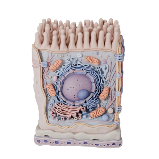
Plant cells have a cellulose cell wall, chloroplasts, a large central vacuole and plasmodesmata. Animal cells have none of these; they have centrioles, and rely on lysosomes and an extracellular matrix instead of a wall. But the shared list is longer than the different one, and this is where misconceptions cluster. Both have a nucleus, endoplasmic reticulum, Golgi apparatus, ribosomes, a cytoskeleton and a plasma membrane. Most importantly, plant cells absolutely do have mitochondria and do perform aerobic respiration — photosynthesis makes glucose, but the plant still has to respire that glucose to get usable ATP, and it does so continuously, day and night. A plant cell in the dark behaves metabolically much like an animal cell.
What to look for in the 3D model
Rotate to a corner-on view first so you can see the flat faces the wall imposes — plant cells are polyhedral rather than round precisely because rigid walls meet at angles. Look for the dense green bodies distributed through the thin peripheral cytoplasm; those are chloroplasts, and note how they sit near the surface where light reaches them rather than deep in the cell. Then find the boundary of the central vacuole and register how little room is left for everything else.