Why the nucleus has multiple lobes
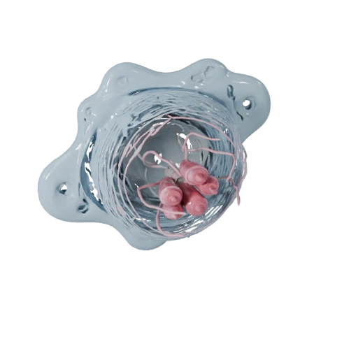
The defining feature visible in the model is a nucleus divided into three to five lobes joined by thin strands of chromatin — which is why neutrophils are also called polymorphonuclear leukocytes. This is not decorative. A neutrophil has to squeeze through gaps between endothelial cells barely wider than a fraction of its own diameter, and a single large rigid nucleus would physically prevent that. A segmented nucleus deforms and flows through in sequence. Lobe count also carries diagnostic information: immature neutrophils released early from marrow have an unsegmented, curved nucleus and are called band cells, while abnormally hypersegmented nuclei with six or more lobes point toward vitamin B12 or folate deficiency.
Three kinds of granules, deployed in order
The grainy cytoplasm gives granulocytes their name, and neutrophils carry several distinct populations. Primary or azurophilic granules hold the most destructive cargo: myeloperoxidase, defensins and neutrophil elastase. Secondary or specific granules, the most numerous, contain lactoferrin, lysozyme and collagenase. Tertiary or gelatinase granules carry matrix-degrading enzymes that help the cell tunnel through tissue, and secretory vesicles supply membrane proteins needed for adhesion. They are released in a deliberate sequence during migration and killing, with the most damaging contents held back until the cell has reached its target — a control problem the cell has to solve, since these enzymes injure host tissue just as readily as bacteria.
Getting to the infection: rolling, adhesion and diapedesis
Neutrophils reach infection through a well-defined sequence. Inflammatory signals cause endothelial cells near the site to display selectins, which catch passing neutrophils and slow them into a rolling motion along the vessel wall — margination. Chemokine signalling then activates integrins on the neutrophil surface, which bind endothelial ICAM-1 and arrest the cell firmly. The neutrophil flattens, finds a junction between two endothelial cells and squeezes through into the tissue, a step called diapedesis or transmigration. Once outside the vessel it navigates by chemotaxis, crawling up a concentration gradient of signals such as interleukin-8, the complement fragment C5a, leukotriene B4 and bacterial peptides. Each step is a separate potential point of failure, which is why defects in adhesion molecules produce recurrent bacterial infection despite normal neutrophil counts.
Phagocytosis and the respiratory burst
Once in contact with a pathogen, the neutrophil engulfs it. Recognition is greatly improved by opsonins — antibody Fc regions and the complement fragment C3b coating the microbe — which the neutrophil binds through dedicated receptors. Pseudopods extend around the target and fuse, enclosing it in a phagosome, which then fuses with granules to become a phagolysosome. Killing follows two routes. The oxygen-dependent route is the respiratory burst: the enzyme NADPH oxidase assembles on the phagosome membrane and pumps out superoxide, which is converted to hydrogen peroxide and then, via myeloperoxidase acting on chloride, to hypochlorous acid — chemically, bleach. The oxygen-independent route relies on granule enzymes and defensins directly. The importance of the oxidative route is demonstrated by chronic granulomatous disease, where a defective NADPH oxidase leaves patients vulnerable to specific catalase-positive organisms despite otherwise normal immunity.
Neutrophil extracellular traps, and why pus exists
Neutrophils have a final option. In a process called NETosis they extrude their own chromatin as a web of DNA studded with granule proteins — a neutrophil extracellular trap — which physically snares bacteria outside the cell and concentrates antimicrobial enzymes around them. The cell dies doing it. This is one reason inflammation is so often destructive to host tissue: the same enzymes and oxidants that kill bacteria damage whatever is nearby, and the accumulated remains of spent neutrophils, digested tissue and microbes is what pus physically is.
Reading neutrophils in a blood count
Neutrophils make up roughly forty to seventy percent of circulating white cells in adults, more than all other types combined. They are also strikingly short-lived, surviving only hours to a day or so in circulation, which means the marrow must produce them continuously. That turnover is what makes the differential count so clinically useful. Acute bacterial infection drives the marrow to release immature forms, raising the band cell proportion — a left shift. Neutropenia, and specifically a low absolute neutrophil count, is the parameter that determines infection risk after chemotherapy. The other four types complete the picture: lymphocytes, monocytes, eosinophils and basophils, in descending order of abundance.
What to look for in the 3D model
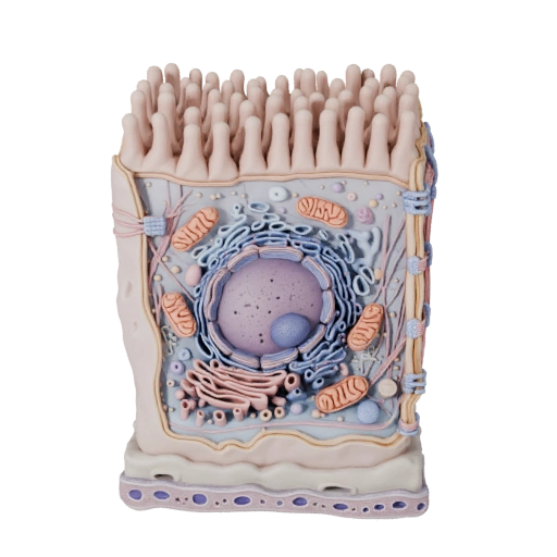
Rotate slowly and watch the outline change — the surface is irregular and lobed rather than smoothly spherical, which is what a cell built to deform looks like when it is not confined to a vessel. Look for the constrictions marking the divisions between nuclear lobes, and note the texture of the cytoplasm where granules are densest. Compare the overall shape to the epithelial cell model: one is built to hold a fixed position in a sheet, the other to change shape constantly.