Three functional zones
Structurally a neuron divides into input, integration and output. Dendrites and the cell body receive signals. The axon hillock integrates them and decides whether to fire. The axon and its terminals carry and transmit the result. This maps onto a clean division of electrical behaviour too: dendrites and soma handle graded, decaying potentials that vary continuously in size, while the axon carries all-or-none action potentials that are identical every time. Information in an axon is therefore encoded in firing frequency, not amplitude — a point worth internalising, because it explains why a stronger stimulus produces faster firing rather than bigger spikes.
Dendrites: receiving and summing thousands of inputs
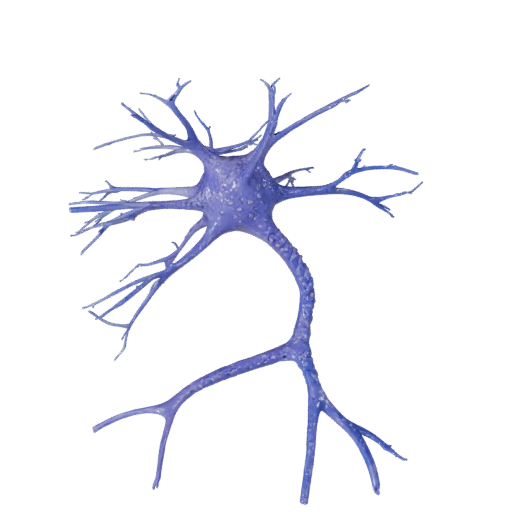
The branching tree occupying most of the model's upper half is the dendritic field, and a single cortical neuron may carry thousands of synaptic contacts across it, many on small protrusions called dendritic spines. Each incoming synapse produces a small local voltage change — excitatory postsynaptic potentials that push toward threshold, inhibitory ones that pull away. These are graded and they decay as they spread, so a distant input arrives at the soma weaker than a nearby one. The neuron adds them together by spatial summation (many inputs at once in different places) and temporal summation (repeated inputs at the same place in quick succession). The sum, not any single input, determines the outcome.
The axon hillock, where the decision happens
Where the soma narrows into the axon there is a region with the highest density of voltage-gated sodium channels anywhere on the cell. This makes the axon hillock the lowest-threshold point on the neuron and therefore the trigger zone: if summed input depolarises the membrane past roughly negative fifty-five millivolts here, sodium channels open en masse and an action potential fires. Below threshold, nothing happens at all. Above it, the spike is always the same size. That all-or-none behaviour is why the hillock is often called the neuron's decision point.
Myelin and saltatory conduction
The segmented sheath along the axon is myelin, and it is not part of the neuron itself — it is wrapped around it by other cells. In the peripheral nervous system each Schwann cell forms one segment; in the central nervous system a single oligodendrocyte myelinates segments on many different axons. Myelin works by simultaneously raising membrane resistance and lowering capacitance, so current travels further down the inside of the axon before leaking out. Between segments are short bare gaps, the nodes of Ranvier, packed with voltage-gated sodium channels. The action potential regenerates only at the nodes and effectively jumps between them, a mechanism called saltatory conduction. The payoff is enormous: a large myelinated axon conducts at well over a hundred metres per second, while an unmyelinated one of similar diameter manages around one. It also explains the clinical picture when myelin is attacked, as in multiple sclerosis — the axon survives but conduction slows or fails.
The synapse: electrical signal to chemical signal and back
At the swollen ends of the model's lower branches are the synaptic terminals. When an action potential arrives, voltage-gated calcium channels open and calcium floods in. That calcium is the trigger for vesicles loaded with neurotransmitter to fuse with the presynaptic membrane via SNARE proteins and release their contents into the synaptic cleft. The transmitter diffuses across, binds receptors on the postsynaptic cell, and produces the graded potential that becomes that cell's input. Signalling is then terminated by reuptake into the presynaptic terminal, enzymatic breakdown in the cleft, or simple diffusion — which matters pharmacologically, since a large share of psychiatric and neurological drugs act on exactly these steps.
The resting membrane potential it all starts from
At rest the inside of a neuron sits around negative seventy millivolts relative to outside. Two things maintain it. Sodium-potassium ATPase pumps three sodium ions out for every two potassium ions in, which both builds the gradients and contributes a small direct negativity. More importantly, the resting membrane is far more permeable to potassium than to sodium because potassium leak channels are open, so the resting potential sits close to potassium's equilibrium potential. Every action potential is a brief, controlled collapse and restoration of this arrangement.
What to look for in the 3D model
Rotate until the axon points away from you and note how completely different the two ends of the cell are — the dense branching input field versus the single long output cable. Follow the axon and look for the periodic constrictions where the myelin segments meet: those are the nodes, and they are the only places the signal actually regenerates. Then zoom to the terminal ends and notice the swelling, which is where vesicles cluster.